Muscles Anterior Full Body Diagram - Human Muscle Anatomy Quiz. More often they work in groups to produce precise movements. Have a product modelling and rendering project?. Skeletal muscles rarely work by themselves to achieve movements in the body. This is a table of muscles of the human anatomy. It is long and thin, running across the thigh in a inferomedial direction.
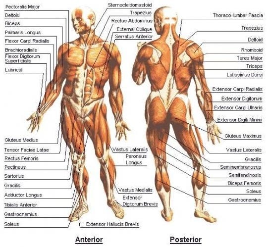
The serratus anterior originates via eight muscular slips that attach to the side of your ribs. Arm anterior 3d illustration project. The scalenus anterior muscle is an important landmark in the neck and is used to find the supraclavicular triangle, which is located near the. Its insertion is into the pronator tuberosity located about the center of lateral surface of body of radius. Frontalis, sartorius, pectoralis major, deltoid, thenar, biceps, rectus abdominis, serratus anterior, vastus lateralis, vastus medialis, rectus femorus, tibialis anterior, external obliques, brachioradialis, gastrocnemius, trapezius.
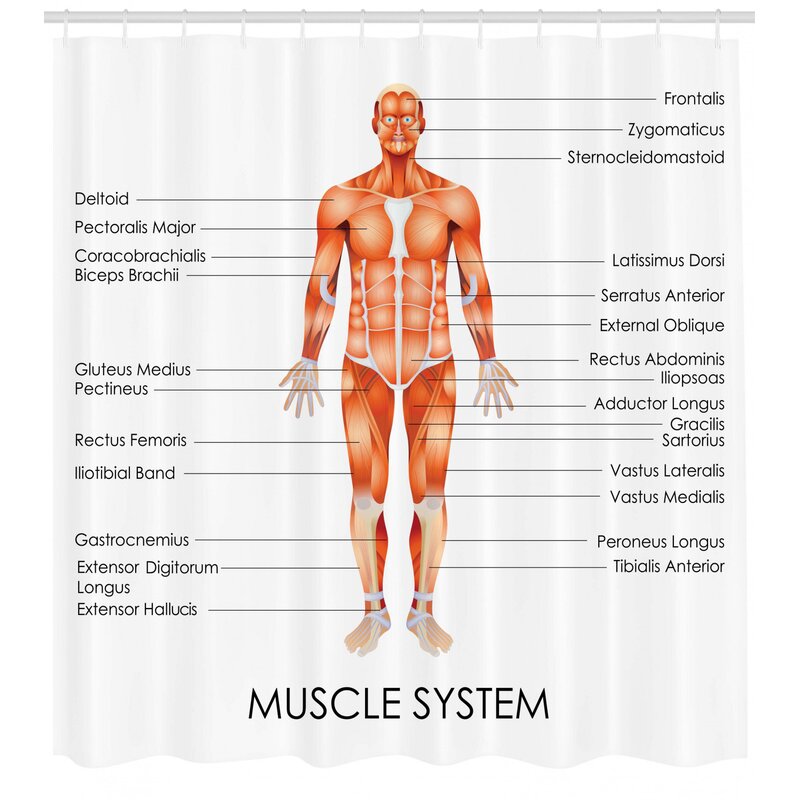
Ambesonne Human Anatomy Muscle System Diagram Of Man Body Features Biological Elements Medical Heath Image Single Shower Curtain Wayfair from secure.img1-fg.wfcdn.com Have a product modelling and rendering project?. In all its forms, it makes up nearly half of the. The human muscular system is complex and has many functions in the body. Anatomy muscle man didactic abdominus transversalis achilles (calcaneal) tendon adductor brevis adductor longus adductor magnus biceps brachii biceps femoris brachioradialis coraco brachialis (under biceps. Anatomical board, anatomical body, human skeleton, anatomy of human bony system, surface anatomy, body shapes, posterior view, full body. More often they work in groups to produce precise movements. Learn faster with these free muscle labeling diagrams. The sartorius is the longest muscle in the body.
Produce wrist and/or finger flexion.
Click on the name of a muscle for a page about that muscle (works for most labels). This muscle diagram is interactive: The serratus anterior muscle attaches your scapula (shoulder blade) to your rib cage and is anatomy. The main framework of the body is covered by muscles, whose function is to permit movement. Get in touch with us today! Produce wrist and/or finger flexion. More often they work in groups to produce precise movements. Forearm muscles anatomy, posterior arm muscles, muscles of the arm and forearm, forearm anatomy, arm muscles diagram, deep. On the next diagram we will indicate the intermediate layer of anterior compartment of forearm. The scalenus anterior (also known as anterior scalene) is a neck muscle and known as the key structure for the thoracic inlet as it is an important anatomical landmark. (1990) principle superficial skeletal muscles. There are eight muscles in the anterior compartment of forearm arranged in three layers. Muscles, connected to bones or internal but muscle is also the dominant tissue in the heart and in the walls of other hollow organs of the body.
Anterior muscles in the body. More often they work in groups to produce precise movements. Get in touch with us today! Anatomy of the human body. Produce wrist and/or finger flexion.
Muscles On The Inside Skin Hair And Nails On The Outside Key Body Parts By Sam Kneller The Explanation Medium from miro.medium.com 353 x 599 photo description: Learn vocabulary, terms and more with flashcards, games and other study tools. Skeletal muscles rarely work by themselves to achieve movements in the body. The thoracic muscles attach on the anterior and lateral regions of the thorax, or rib cage. Muscles of the anterior compartment of the forearm. Arm anterior 3d illustration project. This muscle diagram is interactive: Their main function is contractibility.
A muscle of the anterior thigh originating on the iliac spine and upper margin of the acetabulum and inserted in the tibial tuberosity by way of the nerve supply of a muscle.
The thoracic muscles attach on the anterior and lateral regions of the thorax, or rib cage. Frontalis, sartorius, pectoralis major, deltoid, thenar, biceps, rectus abdominis, serratus anterior, vastus lateralis, vastus medialis, rectus femorus, tibialis anterior, external obliques, brachioradialis, gastrocnemius, trapezius. 353 x 599 photo description: (1990) principle superficial skeletal muscles. Muscles, connected to bones or internal but muscle is also the dominant tissue in the heart and in the walls of other hollow organs of the body. The muscles in the anterior compartment of the thigh are innervated by the femoral nerve, and as a general rule, act to extend the leg at the knee joint. There is one on each side of your body. You have two serratus anterior muscles; This is a table of muscles of the human anatomy. The human muscular system is complex and has many functions in the body. The muscular system consists of various types of muscle that each play a crucial role in the function of the body. It is long and thin, running across the thigh in a inferomedial direction. Anatomy of the human body.
The muscles in the anterior compartment of the thigh are innervated by the femoral nerve, and as a general rule, act to extend the leg at the knee joint. You have two serratus anterior muscles; There is one on each side of your body. Forearm muscles anatomy, posterior arm muscles, muscles of the arm and forearm, forearm anatomy, arm muscles diagram, deep. Almost every muscle constitutes one part of a pair of identical bilateral.
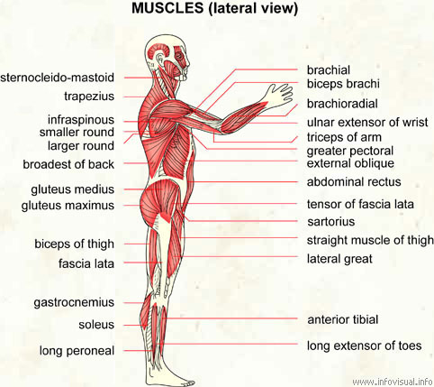
Muscles Lateral View Visual Dictionary from infovisual.info Arm anterior 3d illustration project. It is long and thin, running across the thigh in a inferomedial direction. This muscle diagram is interactive: The human muscular system is complex and has many functions in the body. The serratus anterior muscle attaches your scapula (shoulder blade) to your rib cage and is anatomy. These include mobility, stability, posture, circulation, digestion, and more. Start studying anterior muscles full body. Muscles of the anterior compartment of the forearm.
Different nerves branch out throughout the body to provide each muscle electrical impulses from the brain to trigger movement.
Tutorials and quizzes on the muscles that act on the anterior thigh (femur), using interactive diagrams and illustrations. Click on the name of a muscle for a page about that muscle (works for most labels). (1990) principle superficial skeletal muscles. The longus colli is situated on the anterior surface of the vertebral column, between the atlas and the third thoracic vertebra. This is a table of muscles of the human anatomy. The muscles labelled in the anterior muscles diagram shown above are listed in bold in the following table Almost every muscle constitutes one part of a pair of identical bilateral. Their main function is contractibility. Anterior muscles in the body. In all its forms, it makes up nearly half of the. Anatomical board, anatomical body, human skeleton, anatomy of human bony system, surface anatomy, body shapes, posterior view, full body. Arm anterior muscles labeled 3d illustration. Muscles allow a person to move.